
PET is a powerful imaging modality, functional in nature, which uses positron emitting isotopes attached to specific molecules to provide exceptionally sensitive assays of a wide range of biological processes. Its principal drawback is relatively poor spatial resolution, making unambiguous localization of signal extremely difficult in many cases. Magnetic resonance imaging (MRI), on the other hand, provides exquisite high-resolution anatomical information, as well access to volume specific chemical and physical information (e.g. metabolite concentrations). For these reasons, PET and MR techniques are largely complementary and merging of these two modalities in the study of experimental animal models would allow us to exploit, in a synergistic fashion, the strengths of both techniques. We are currently developing a high resolution, high sensitivity multi-ring PET scanner integrated in a 7 Tesla/30 cm small animal MRI system. The PET system was designed to minimally interfere with the MRI, and to itself be insensitive to outside interference (Fig. 1).
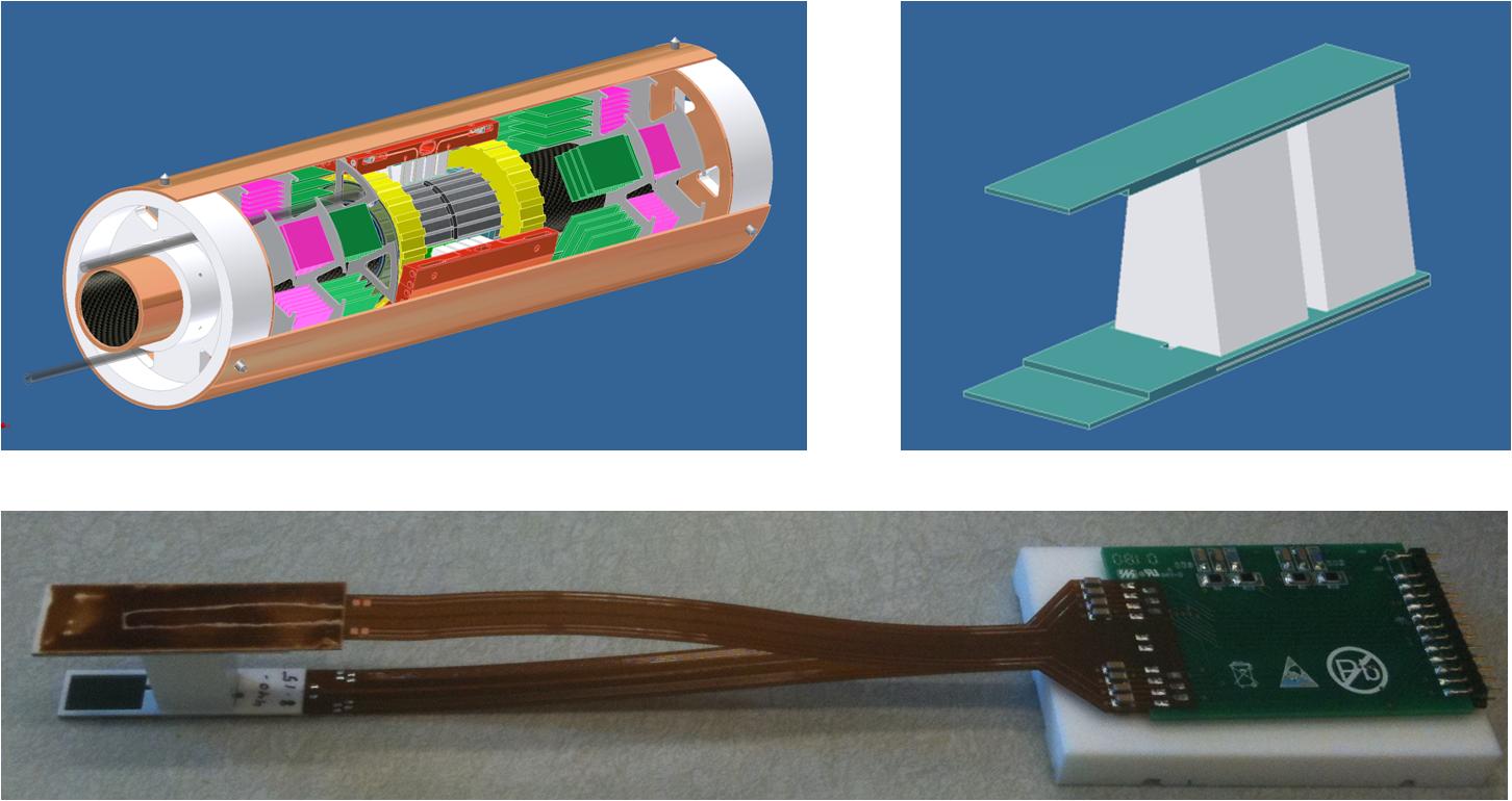
Fig. 1: top:Schematic illustration of the new generation PET insert and the used tapered detector blocks.
bottom: image of an assembled detector module with preamplifier stage.
We have obtained simultaneous imaging results in phantoms and in vivo, with an initial prototype system. We are developing the next generation with higher resolution, higher sensitivity and better robustness for simultaneous PET/MR studies (Fig. 2).
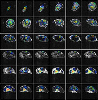
Fig. 2: Combined PET and MRI images of a mouse injected with radioactive F-18