
Although positron emission tomography (PET) is the most sensitive molecular imaging tool available for human imaging, current scanners are constrained by the short scanner length (~20 cm). This imposes several limits on the use of PET: long scan times are needed to produce whole-body images with sufficient signal-to-noise, and it is impossible to place all organs and tissues in the field-of-view at once.
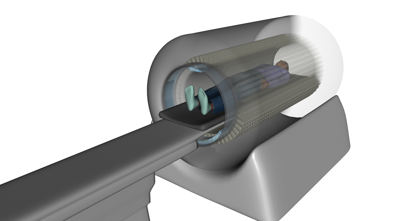
Figure 1. Artistic rendering of the EXPLORER scanner showing full coverage of the patient.
To overcome these constraints, we aim to build the EXPLORER scanner: a high sensitivity, total-body PET system with a 2 meter axial field-of-view. (explorer.ucdavis.edu). This is a large collaborative effort across several academic research groups as well as a partnership with industry. The scanner geometry (Figure 1) places all the organs in the field-of-view and drastically increases the efficiency of capturing the PET signal. Ultimately, this can lead to improved image quality (improved lesion detection and quantification), shorter scan times (minimizing motion artifacts), and reduced radiation dose (crucial for pediatrics and longitudinal imaging). These advances have the potential to produce a paradigm shift in how PET is used and to expand our understanding of human biology in health and disease.
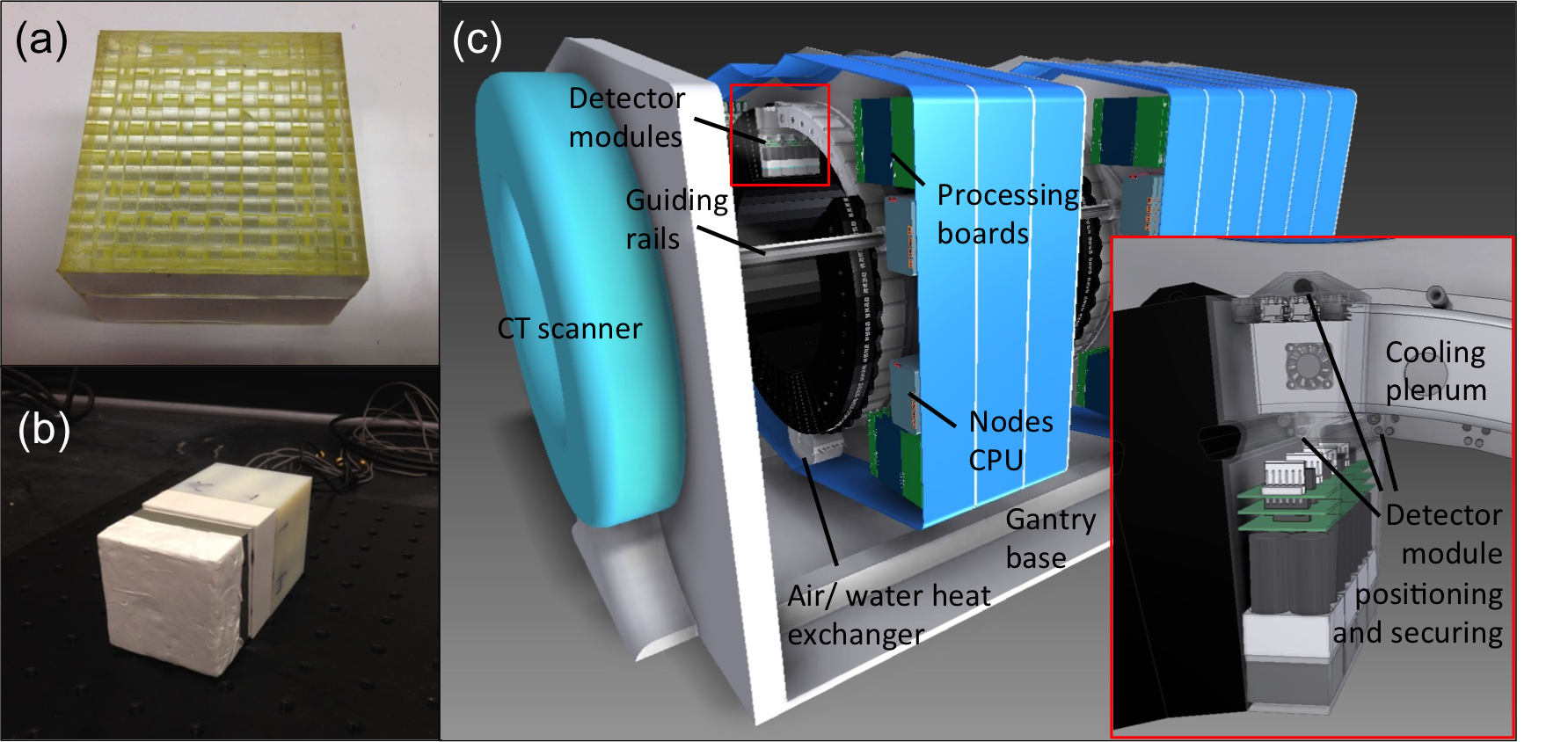
Figure 2. a) Phosphor-coated scintillator array and b) Assembled detector module. c) Scanner gantry design schematic.
Our laboratory is involved in several areas of the project including simulations, detector development, scanner gantry design, electronics and data handling techniques. To accommodate the scanner’s large field-of-view, we have implemented phosphor coating to gain depth-of-interaction information in a time-of-flight commercial PET detector. We are currently extending this work in a custom detector module (Figure 2a-b) with advanced signal processing techniques as well as investigating SiPM-based detectors. No PET scanner of this scale has been built; therefore, we are actively working on a scanner gantry design to handle the enormous scale of the system and heat dissipation from the detectors and electronics (Figure 2c). Along with this, we are developing data handling methods to sort and analyze the data stream output from the electronics (up to 10 Gb/sec) in order to find coincident event pairs, etc.
Lastly, we are building a scaled-down version of the EXPLORER scanner for non-human primate imaging. This scanner is based on a standard clinical system that is reconfigured to suit the primate anatomy (smaller bore, longer axial length). We will use this scanner to prototype potential imaging applications in primates before the full EXPLORER system is completed.
References:
1. Poon, JK; et al. Optimal whole-body PET scanner configurations for different volumes of LSO scintillator: a simulation study. Phys. Med. Biol. 57(13), pp. 4077, 2012.