A high resolution and high sensitivity dedicated mouse brain PET scanner
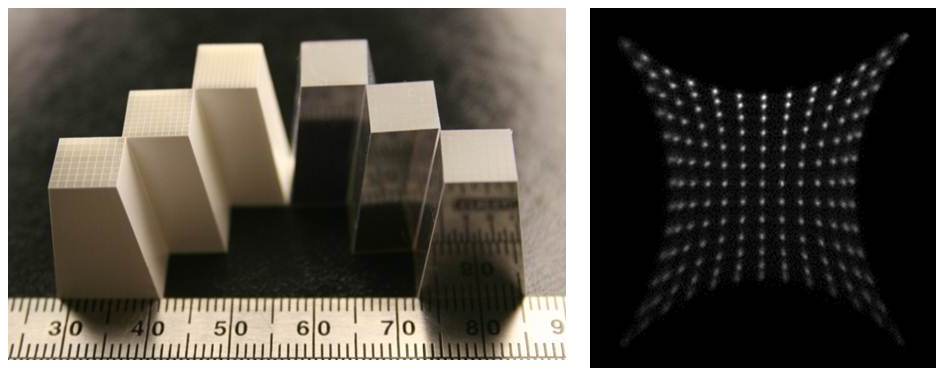
The performance of available small animal PET scanners continues to be detector rather than physics limited. Significant improvements in the sensitivity and spatial resolution of a small animal PET scanner would not only improve the quantification of signals that we are currently able to visualize (by reducing partial volume effects and increasing signal-to-noise) but would also open up important new applications that are beyond the reach of contemporary animal PET systems. Our approach is to develop depth-encoding PET detectors that simultaneously exhibit high spatial resolution and high efficiency by using dual-ended readout of high-efficiency and finely pixelated scintillator arrays with position sensitive avalanche photodiodes (PSAPDs). We have successfully developed both rectangular and tapered lutetium oxyorthosilicate (LSO) arrays with crystal sizes down to 0.5 mm, very high efficiency (20 mm thick detectors) and a depth of interaction (DOI) resolution of ~ 2 mm (Fig. 1).

Fig. 1. (Left) Six tapered LSO arrays with different reflector and crystal size, (right) flood histogram of a tapered LSO array with crystal size of 0.51×0.51 mm 2 at the small end, 0.51×0.91 mm2 at the large end and 20 mm long.
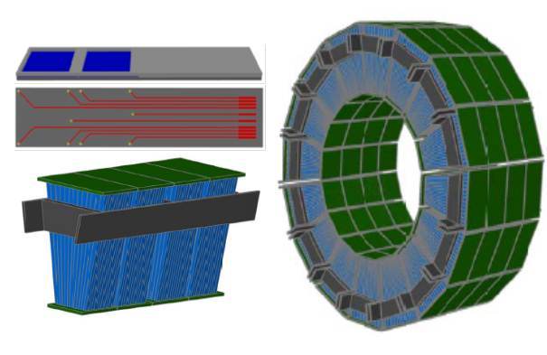
We are currently working on developing a preclinical PET scanner designed for molecular imaging studies in the mouse brain. The scanner will use tapered depth-encoding PET detectors that simultaneously exhibit high spatial resolution and high efficiency. This will open up new opportunities for quantitative molecular imaging studies of development and aging, studies of murine disease models (Alzheimer’s disease, Parkinson’s disease, stroke, and neurodevelopmental disorders to name but a few), and monitoring of novel therapeutic strategies based on small molecules, peptides, nanoparticles, gene therapies or cellular therapies.

FIg. 2: (Top-left) New type of PSAPDs, (bottom-left) a detector cassette with four depth encoding detectors, (right) all detectors of the proposed dedicated mouse brain PET scanner.
Reference:
1) Yang Y, Wu Y, Qi J, James SS, Du H, Dokhale PA, Shah KS, Farrell R, Cherry SR. A prototype PET scanner with DOI-encoding detectors. J Nucl Med 2008; 49: 1132-1140.
2) Yang YF, St James S, Wu YB, Du H, Qi J, Farrell R, Dokhale PA, Shah KS, Vaigneur K, Cherry SR. Tapered LSO arrays for small animal PET. Phys Med Biol 2011; 56:139-153.
